Rotary
Biomechanics: Reality or Future?
Jesus Djalma Pécora
Alexandre Capelli
Fábio Heredia Seixas
Melissa Andréia Marchesan
Danilo Mathias Zanello Guerisoli
A
major concern regarding rotary instruments is their unexpected fracture while
in operation. It can occur without any visible sign of permanent deformation,
and the two most important causes of fractures are torsional and flexural
failures (SERENE et al., 1995). Torsional fractures occur when the tip or any
other part of the instrument engages the root canal while the other part still
rotates. Flexural failures are those caused by metal fatigue, when the
instrument is used in a canal with a small curve radius. The limit of
flexibility of the instrument is exceeded, thus resulting in fracture by cyclic
fatigue (PRUETT et al., 1997; LOPES et al., 1999).
New
procedures, able to reduce the occurrence of instrument fractures, have been
researched. Many techniques of root canal preparation, with different
instruments and engines, have been recommended by authors and manufacturers
(LEONARDO & LEONARDO, 2002).
This
study suggests a new method for root canal preparation with rotary Ni-Ti
instruments that can reduce the rate of fractures, using files from any
manufacturer and either electric or pneumatic engines.
THE FREE-TIP PREPARATION TECHNIQUE
First considerations
Perform
access surgery, irrigate pulp chamber and root canals copiously with sodium
hypochlorite, leaving it inundated with the solution. Initial exploration of
the orifices must be performed with an endodontic explorer or manual files of
compatible diameter.
Nickel-titanium
rotary instruments of any manufacturer can be used. It is possible to combine
different brands, choosing the most suitable for each step of treatment.
The
engine can be either electric or pneumatic, from any manufacturer. This
technique suggests the use with a speed between 250 and 350 rpm.
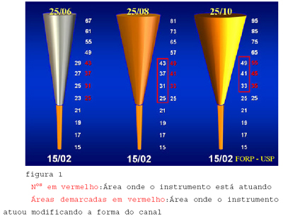
Cervical preparation
A
Ni-Ti #25 taper .06 instrument must be attached to the handpiece and the engine
speed must be set between 250 and 350 rpm.
Start
the motor and using a smooth pecking motion, initiate the instrumentation of
the root canal following its long axis. Avoid eccentric movements of the file,
neither try to force it laterally.
Continuing
with the instrument inside the root canal longer than the necessary to reach
working length (around 2/3 of the total length at this stage) must be avoided.
At 300 rpm, the instrument will perform 5 turns per second around its axis.
Irrigate
with sodium hypochlorite and alternate it with EDTA. Tissue dissolution is
directly proportional to sodium hypochlorite concentration.
A
new Ni-Ti instrument must be chosen, with greater taper (.08, .10 or .12) than
the previously used (Orifice Shaper,
Dentsply-Maillefer; GT Accessories,
Dentsply-Maillefer; Flare, Quantec).
Tip diameter (D1) must be about 25 – 40 (Figure
1).
Apical preparation
Following
cervical preparation, a minor taper (thus, more flexible) instrument must be
chosen for apical preparation (15/.04 or 20/.02). As a result, the file will
operate without cervical interferences and will access curves due to its
flexibility, reaching the temporary working length. The working length must be
then determined by an X-ray.
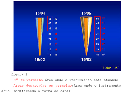
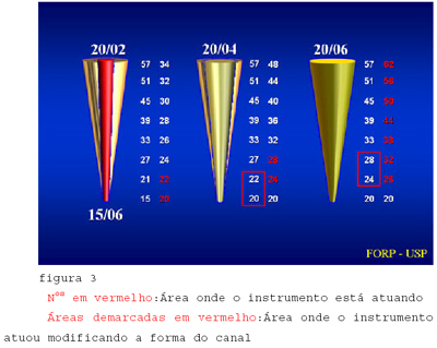
The
root canal preparation must be continued with the following instruments:
20/.02; 20/.04; 25/.04 or 15/.04; 15/.06; 20/.04 and 25/.04 until working
length is reached.
If
any instrument does not reach the working length, irrigate the root canal with
sodium hypochlorite and use one or two instruments with greater taper than the
used previously. Then try to use the instrument that did not reach the working
length. Always remember to irrigate copiously the root canal with sodium
hypochlorite (Figures 2 and 3).
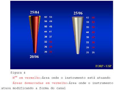
Finalization
Choose
an instrument with intermediary taper than the previously used for apical
preparation and tip diameter equal or smaller than that. This instrument will smooth the
irregularities and promote a conical and continuous shape of the root canal (Figure
4).
The
files used at this stage are:
Discussion
There
are many reason for instrument fractures. One of them is the radius of the
curvature and its location (PRUETT, 1997). The smaller the radius, higher will
be the stress submitted to the instrument (LOPES, 1999). Clinically, the small
radius curves are located at the apical third of teeth, which favors fractures
near the tip of the file. Instruments with greater taper are more prone to
fractures when used in sharp curves.
Another
factor that raises the incidence of fractures is the increase of pressure
applied by the operator to the apex (BLUM et al., 1999). The speed of rotation
of the instruments was also reported as directly responsible for fractures
(DIETZ et al., 2000).
Pressure
control and movement applied to instruments, as well as the use of engines
especially developed for this aim with speed reduction, help to avoid
instrument fractures.
The
risk of fractures is always increased when dentine cutting is performed with
the tip of the instrument (BLUM et al. 1999). With excessive apical pressure
and high torque, the instrument ruptures.
The
free-tip preparation technique aims to prepare the root canal with areas of the
instrument with greater taper first, leaving the tip free. This reduces
dramatically the occurrence of fractures and was described in a similar way by
other authors (McSPADDEN, 1996; BASSI apud LEONARDO & LEONARDO, 2002). The
majority of instruments break at their most fragile portion, i.e., the tip or
near it.
In
order to avoid such problem, preparation can be initiated with a smaller taper
instrument and facilitate the way for the next file, which will have its tip
working freely inside the canal, acting just as a guide. Thus, the root canal
will be prepared in a crown-down way. In order to reach the working length, the
instrument must prepare the cervical portion of the canal first, enlarging it
up to the apex. The areas of the instrument with greater metallic structure
will receive the forces during biomechanical preparation.
The
knowledge from the manual files does not apply to the rotary files, since
stainless steel is very different from nickel titanium in metallurgic aspects.
Thus, the files have a different mechanical behavior.
Conclusion
Root
canal preparation with Ni-Ti rotary files is a worldwide reality, including
Brazil. This can be confirmed by the number of courses focusing this new
technology and its introduction in many universities as part of the discipline
of Endodontics.
References
1.
AHLQUIST, M.; HENNINGSSON, O.;
HULTENBY, K. & OHLIN, J. The effectiveness of manual and rotary techniques
in the cleaning of root canals: a scanning electron microscopy study. I Endod J,
v. 34, p. 533-537, 2001
2.
BLUM, J.Y.; COHEN, A.; MACHTOU,
P.; MICALLEF, J.P. Analysis of forces developed during mechanical preparation of
extracted teeth using ProFile NiTi rotary instruments. I Endod J, V.32, p.
24-31, 1999
3.
BORTNIK, K.L.; STEIMAN, H.R.;
RUSKIN, A. Comparison of nickel-titanium file distortion using electric and
air-driven handpiece. J Endod v.27, n.1, p.57-59, 2001
4.
BUCHANAN, L. S. The
standardized-taper root canal preparation - Part 1. Concepts for variably
tapered shaping instruments. Int Endod J,
v.33, p.516-529, 2000.
5.
BUCHANAN, L. S. The
standardized-taper root canal preparation - Part 2. File selection and safe
handpiece - driven file use. Int Endod J,
v.34, p.63-71, 2001.
6.
DEUS, Q.D. de. Endodontia.
5a ed., Medsi, Rio de Janeiro, 1992. 695p.
7.
DIETZ, D.B.; DI FIORE, P.M.;
BAHCALL, J.K.; LAUTENSCHLAGER, E.P. Effect of rotational spesd on the breakage
of Nickel-Titanium rotary files. J Endod, v.25, n.2, p. 68-71, 2000
8.
HÜLSMANN, M.; SCHADE, M.; SCHÄFERS,
F. A comparative study of root canal preparation with HERO 642 and Quantec SC
rotary Ni-Ti instruments. I End Journal,
v.34, n.5, p.538-546, 2001.
9.
LEONARDO, M.R. LEONARDO, R.T. Sistemas Rotatórios em
Endodontia-Instrumentos de Níquel-Titânio, Artes Médicas, 2002.
10.
LOPES, H.P., SIQUEIRA JR, F., ELIAS, C. Instrumentos
Endodônticos. In: LOPES, H.P., SIQUEIRA JR, F. Endodontia. Biologia e Técnica. Rio de Janeiro: Medsi, p.279-318,
1999 (a).
11.
LUMLEY, P. J. Cleaning efficacy
of two apical preparation regimens following shaping with hand files of greater
taper. Int Endod J, v.33, p. 262-2, 2000
12.
MacSPADDEN, J.T. Advanced
geometries in endodontic micro files: The rationale Chattanooga, The NT Company
(1996).
13.
PETERS, O. A.; BARBAKOW, F. Effects of irrigation on
debris and smear layer on canal walls prepared by two rotary techniques: a
scanning electron microspic study. J
Endod, v.26, n.1, p.6-10, 2000.
14.
PRUETT, J.P.; CLEMENT, D.J.; CARNES,D.L. Ciclic fatigue
testing of nickel-titanium endodontic instruments. J Endod, v.23, p.77-85, 1997.
15.
SATTAPAN,B.; NERVO, G.J.;
PALAMARA, J.E.A.; MESSER, H.H. Defects in rotary Nickel-titaium files after
clinical use. J Endod, v.26, n.3, p.161-165, 2000
16.
SCHÄFFER, E. Root canal
instruments for manual use: a review. Endod Dent Traumat, v. 13, p. 51-64, 1997.
17.
SERENE, T.P.; ADAMS, J.D.,
SAXENA, A. Nickel-Titanium Instruments:
Applications in endodontics. St. Louis Missouri, USA: Ishiyaku
Euroamerica, Inc., 112p, 1995.
18.
SIQUEIRA, J.F.; ARAÚJO, M.C.;
GARCIA, P.F.; FRAGA, R.C.; DANTAS, C.J. Histological evaluation of the
effectiveness of five instrumentation techniques for cleaning the apical third
of root canals. J Endod, vol.23,
n.8, p.499-502, 1997.
19.
THOMPSON, S. A.; DUMMER, P. M. Shaping ability of
ProFile .04 taper series 29 rotary nickel-titanium instruments in
simulated root canals: Part 1. Int Endod
J, v.30, n.1, p.1-7, 1997a.
20.
THOMPSON, S. A.; DUMMER, P. M. Shaping ability of
ProFile .04 taper series 29 rotary nickel-titanium instruments in
simulated root canals: Part 2. Int Endod
J, v.30, n.1, p.8-15,1997b.
21.
WALIA, H.; BRANTLEY, W.A.;
GERSTEIN, H. – An initial investigation of the bending and torsional
properties of nitinol root canal files. J Endod vol.14, nº. 7, p.346-51,
1988.
22.
YARED,
G.M.; BOU DAGHER & MACHTOU, P. Cyclic fatigue of ProFile rotary instruments
alter clinical use. I
End Journal, v.33, p.204-207, 2000.
23.
YARED,
G.M.; BOU DAGHER & MACHTOU, P. Failure of Profile instruments used with high
and low torque. I
End Journal, v.34, p.471-475, 2001.
24.
YARED,
G.M.; BOU DAGHER & MACHTOU, P. Inflense of rotacional speed, torque and
operator’s proficiency on ProFile failures. I End Journal, v.34,
p.47-53, 2001.